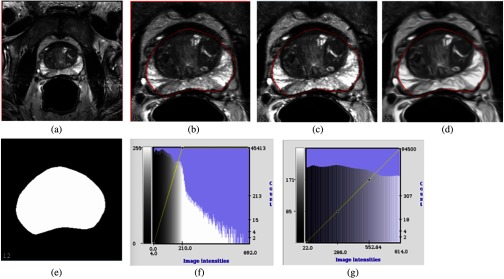
Fig. 6.
An example of MR prostate image preprocessing steps: (a) The original 3-D MRI slice, dimension . (b) The cropped image after 25% region reduction from top, bottom, left, and right. (c) Applying median + IQR scaling to generate the MRI slices before HNN training. (d) CED applies to image after median + IQR scaling. (e) Corresponding prostate binary masks for both MR and CED images. (f) The histogram of the original MRI after cropping. (g) The histogram after applying median + IQR with intensity scaling range from 0 to 1000, lastly rescaled to [0, 255] to fit to the PNG image format for training. After the preprocessing steps, the median + IQR scaling generated MRI slice and the CED image slice with binary mask pairs are mixed together to train one single HNN model.

