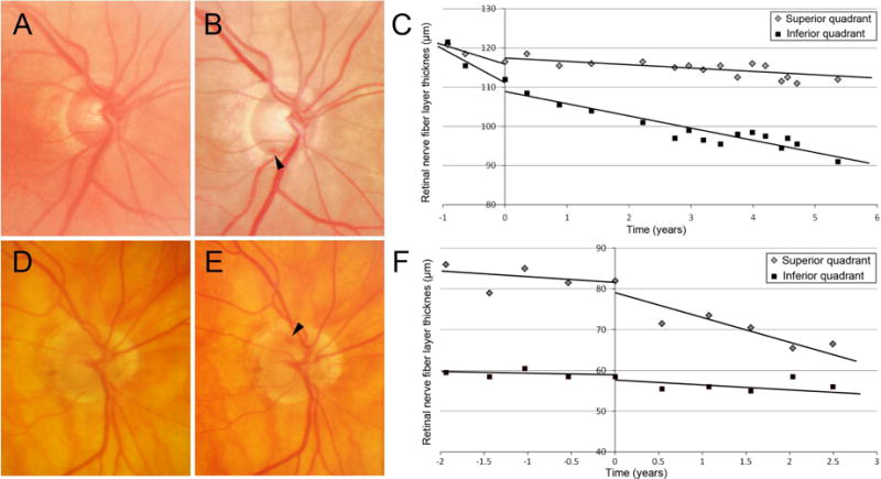
Figure 1.

Examples of eyes with disc hemorrhage. A–C, Optic disc photograph shows no DH at the first examination (A) and inferotemporal DH at the episode of DH (B, time is set as 0). The rate of superior RNFL thinning of −3.25 μm/year before DH decreased to −0.83 μm/year after the episode of DH. The rate of inferior RNFL thinning before and after DH was −4.02 and −2.93 μm/year, respectively. The glaucoma medication was intensified from 1 to 2 eye drops after DH. D–F, Optic disc photograph shows no DH at the first examination (D) and superotemporal DH at the episode of DH (E, time is set as 0). The rate of superior RNFL thinning of −0.49 μm/year increased to −5.04 μm/year after the episode of DH. The rate of inferior RNFL thinning before and after DH was −0.21 and −1.08 μm/year, respectively. The glaucoma medication score was 2, which was not altered after DH. The RNFL slope value was calculated using mixed effects modeling.
