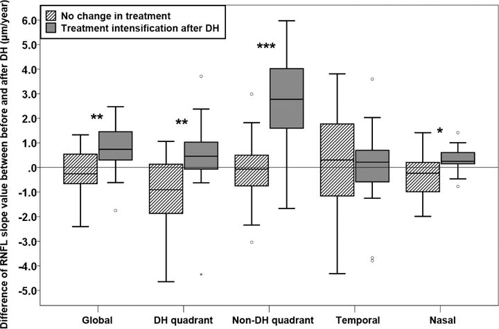
Figure 2.

Difference of global and local retinal nerve fiber layer (RNFL) slopes before and after disc hemorrhage (DH). Positive values indicate that the rate of RNFL thinning became slower after DH compared to before DH. Negative values indicate that the rate of RNFL thinning became faster after DH compared to before DH. In global, DH, non-DH, and nasal quadrants, the rate of RNFL slope values significantly slowed more in eyes with intensified glaucoma treatment compare to eyes without treatment change. It should be noted that the slope became significantly faster (more negative) in DH quadrant in eyes without treatment change. *, **, and *** indicate P < 0.05, < 0.01, and < 0.001, respectively.
