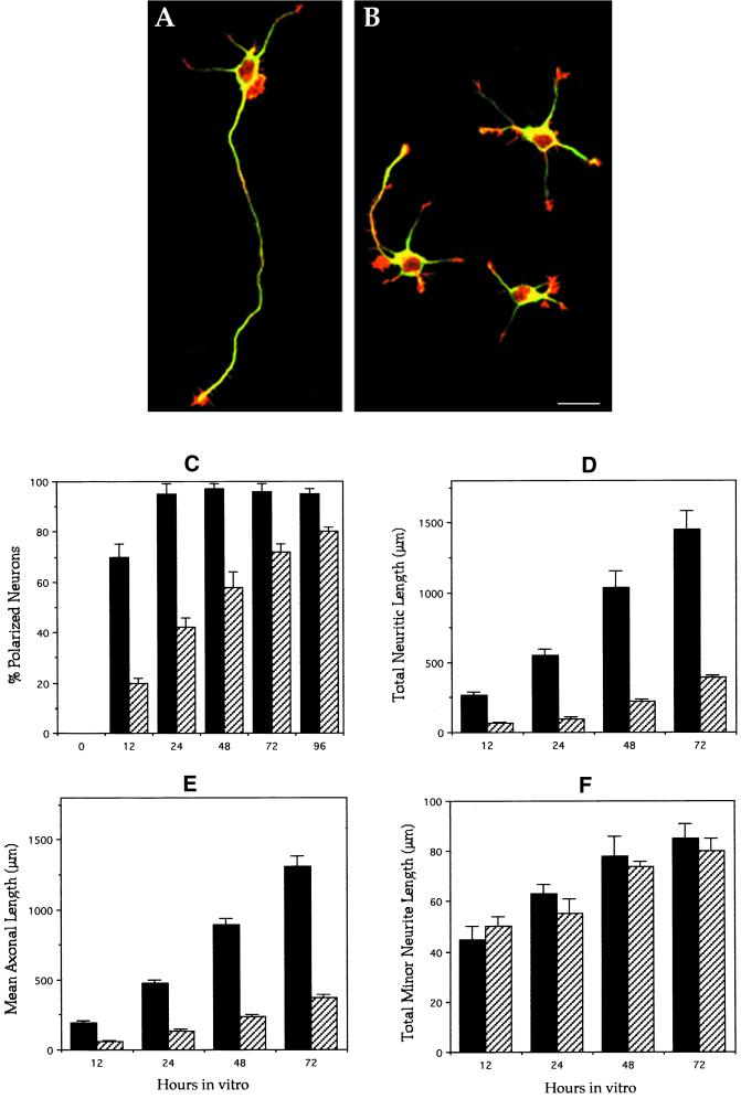
Figure 2.
General morphology and morphometric parameters of control and MAP1B-deficient neurons. (A) Confocal micrograph showing a polarized hippocampal pyramidal neuron from a wild-type animal. The cell that displays one single long axon and several much shorter minor neurites was maintained in culture for 2 d. The cell is double labeled with a mAb against tyrosinated α-tubulin (green) and rhodamine-phalloidin (red). (B) Confocal micrograph showing cultured hippocampal pyramidal neurons from a homozygous (−/−) MAP1B mutant mice. The cells were cultured for 2 d and stained as in B. Note that only one cell is polarized and displays a very short axon-like neurite. Bar, 20 μm. (C) Graph showing the percentage of cells displaying axon-like neurites in cultures from wild-type (▪) and MAP1B-deficient (▨) mice. (D–F) Graphs showing changes in total neuritic length (D), axonal length (E), and total minor neuritic length (F) in hippocampal cell cultures from wild-type (▪) and MAP1B-deficient (▨) mice. Note the significant and selective decrease of axonal length in the MAP1B-deficient neurons. Values represent the mean ± SEM.

