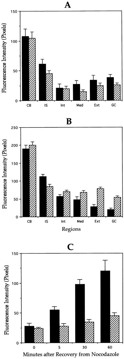
Figure 6.
Quantitation of stable and dynamic microtubules in control and MAP1B-deficient neurons. Quantitative fluorescence measurements of tyrosinated (A) and detyrosinated (B) α-tubulin immunolabeling in cytoskeletal preparations from wild-type (▪) or MAP1B-deficient (▨) cultured hippocampal pyramidal neurons treated with nocodazole for 5 min. Measurements were performed in equivalent regions to those described in Figure 4. (C) Quantitative fluorescence measurements of tyrosinated α-tubulin immunolabeling in cytoskeletal preparations from wild-type (▪) or MAP1B-deficient (▨) cultured hippocampal pyramidal neurons during recovery from nocodazole. For this experiment cultured neurons were treated with nocodazole for 30 min. Fluorescence intensity measurements were performed at the distal axonal segment. A total of 75 cells was analyzed for each experimental condition.

