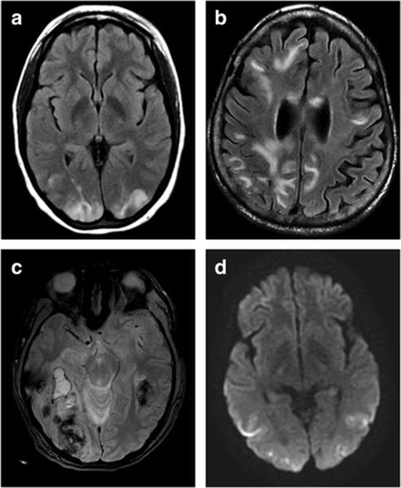
Fig. 1.

Representative imaging findings from 4 cases of PRES: (a) Axial T2 FLAIR image of typical parieto-occipital distribution in a 23-year-old woman without hemorrhage (mRS of 0 at discharge), (b) axial T2 FLAIR image of extensive edema in a 65-year-old woman (mRS of 5 at discharge), (c) axial susceptibility-weighted image of hemorrhage with mass effect in a 36-year-old woman (mRS of 4 at discharge), and (d) axial DWI image showing diffusion restriction in a 25-year-old woman (mRS of 3 at discharge). *mRS: modified Rankin Scale
