
Figure 1.
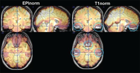
Side‐by‐side comparison of EPInorm and T1norm approaches for a single subject transparently overlaid on the T1 image from that subject. The T1norm process is unable to compensate for distortions throughout the brain, which are not present in the T1 scan (blue circles). [Color figure can be viewed at http://wileyonlinelibrary.com]