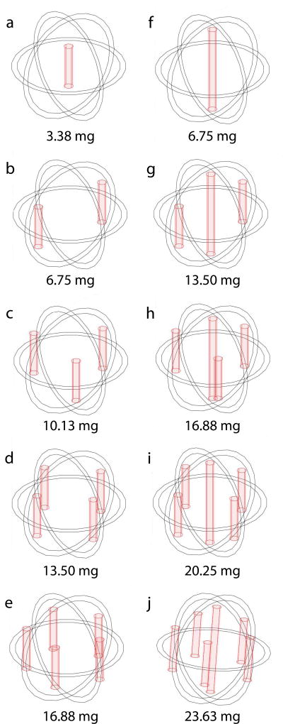
Figure 2.
Multiple implant configurations for simulating drug distribution in tissue with a 2.0 cm diameter tumor (denoted by the outer sphere) with a 1.8 cm diameter ablated tumor core (denoted by the inner sphere). Single central implant with different lengths: (a) 8 mm; (f) 16 mm. Two to five peripheral 8-mm long implants (b–e). Peripheral 8-mm long implants and a central 16-mm long implant (g–j). Total doxorubicin doses are shown below each design.

