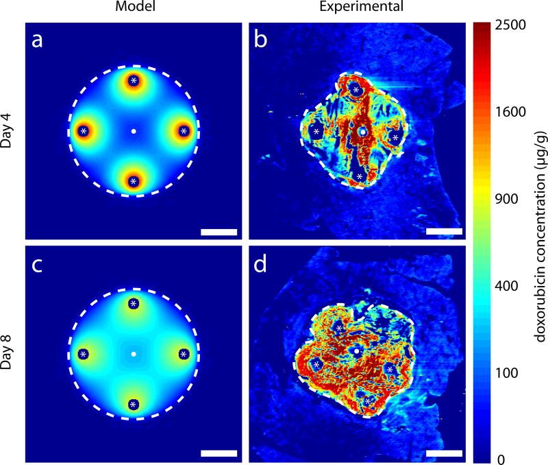
Figure 5.
Simulated drug distribution in ablated normal tissue compared to DOX distribution in images of ablated normal liver from in vivo rabbit experiments with four peripheral implants (located at white asterisks). Ablated regions are within white dashed lines. Thermal ablation source located at white dots. Model simulated DOX distributions on day 4 (a) and day 8 (c). DOX distributions from experimental images on day 4 (b) and day 8 (d). Fixation or preservation artifact in experimental images arose from fracture of brittle ablated tissue during slicing. Scale bars are 5 mm.

