
Figure 1.
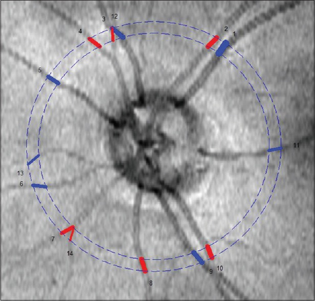
Doppler Fourier domain optical coherence tomography image illustrating the double circular scan pattern around the optic nerve head
Official websites use .gov
A
.gov website belongs to an official
government organization in the United States.
Secure .gov websites use HTTPS
A lock (
) or https:// means you've safely
connected to the .gov website. Share sensitive
information only on official, secure websites.
Doppler Fourier domain optical coherence tomography image illustrating the double circular scan pattern around the optic nerve head