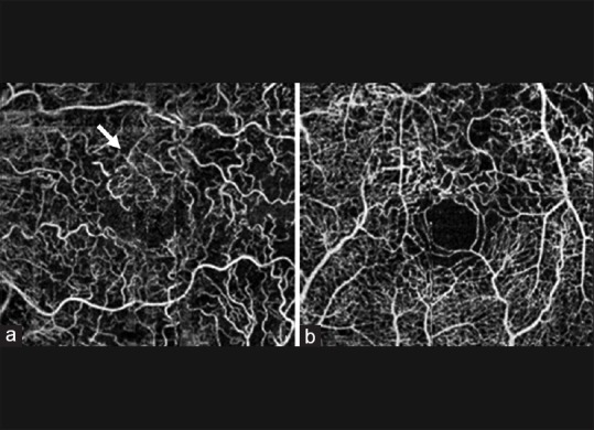
Figure 3.

(a) Unsegmented 3 mm × 3 mm optical coherence tomography angiogram showing collateral formation (white arrows) in an eye with branch retinal vein occlusion. (b) Unsegmented 3 mm × 3 mm optical coherence tomography angiogram showing vascular tortuosity in an eye with central retinal vein occlusion (reprinted from[79])
