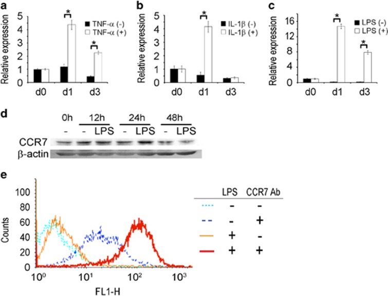
Figure 2.
Inflammatory stimuli increase CCR7 expression in BMMs and osteoclasts. (a–c) The CCR7 mRNA levels in BMMs treated with RANKL in the absence and presence of TNF-α, IL-1β and LPS. BMMs were treated with M-CSF (30 ng ml−1) and RANKL (100 ng ml−1) for 1 day or 3 days with concomitant TNF-α (5 ng ml−1), IL-1β (5 ng ml−1), LPS (5 ng ml−1) or control vehicle. The CCR7 mRNA was measured by real-time PCR. The internal control was the mRNA for 18S rRNA. (d) The CCR7 protein level in BMMs stimulated with LPS. BMMs were treated with LPS (5 ng ml−1) for 12, 24 and 48 h. The whole-cell lysates were used for western blotting. β-Actin was used as a loading control. (e) The cell-surface level of CCR7 protein in BMMs stimulated with LPS. BMMs were treated with LPS (5 ng ml−1) or PBS for 24 h. The cells were collected and subjected without permeabilization to flow cytometry with an anti-CCR7 antibody. The CCR7 antibody was omitted in the controls. BMM, bone marrow-derived macrophages; IL-1β, interleukin-1β LPS, lipopolysaccharide; OA, osteoarthritis; RA, rheumatoid arthritis; TNF-α, tumor necrosis factor-α.

