Figure 4.
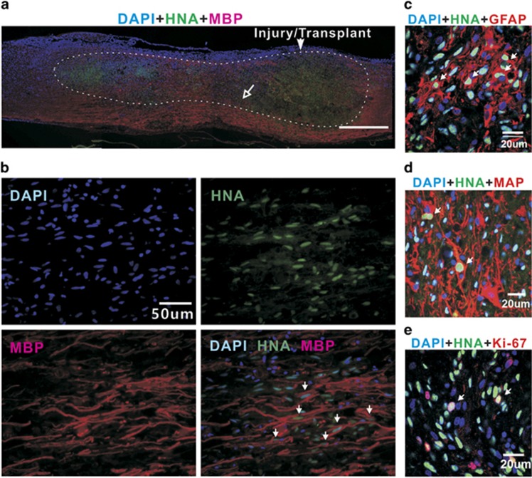
Cells derived from the OPC-like transplants in the injured spinal cord 8 weeks post transplantation. (a) A sagittal section image of the spinal cord illustrating the lesion/transplant site, adjacent area and injury-induced lesion cavity (area within the dotted line) filled with transplants. HNA-positive cells (green) survived and migrated both rostrally and caudally within the injury-induced cavity to distances of at least 5 mm from the transplanted site. Although the majority of HNA-positive cells was located within 1.5–3 mm rostral and caudal to the transplanted site, a larger proportion of cells migrated rostrally. (b) High-magnification images of the small area indicated by the open-headed arrow in a. The survival of oligodendrocytes derived from the transplants was confirmed by triple labeling with DAPI, anti-HNA and anti-MBP antibodies (arrows in photo at the bottom right). (c–e) Coronal section image of the spinal cord, showing other types of surviving cells derived from the transplants (arrows); (c) GFAP-positive cells (red), (d) MAP-positive cells (red) and (e) Ki67-positive cells (red) that are proliferative.

