
FIG 2 .
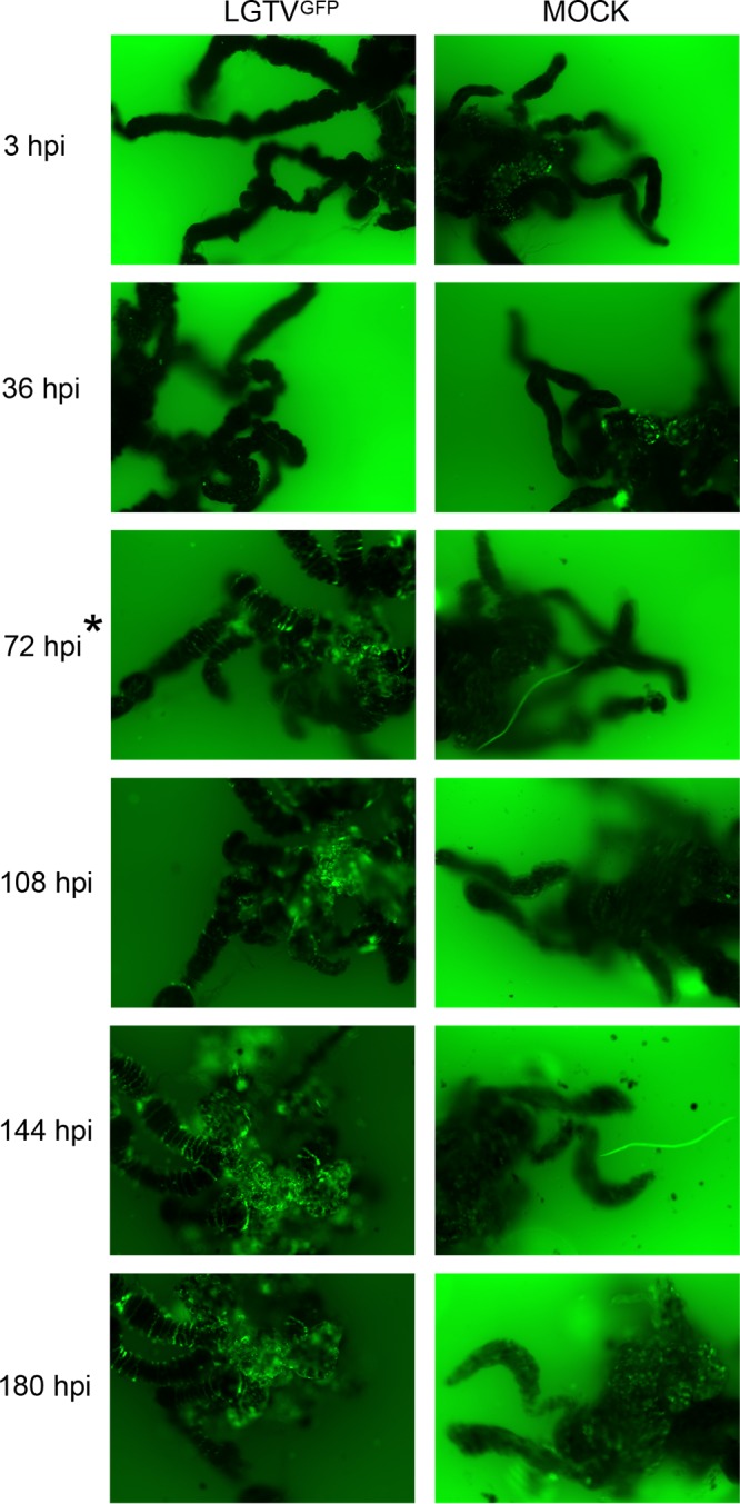
LGTVGFP replication in infected midgut culture. Magnification, ×10 for organs with GFP filter imaging. LGTV expressed GFP, shown in green within the organ. A mock-infected organ was used for comparison, and small amounts of autofluorescence were observed. Serial imaging of the same LGTVGFP-infected and mock-infected midgut organ was performed. The asterisk at 72 hpi denotes the first time point where GFP expression within the LGTVGFP-infected midgut was distinguishable from autofluorescence in the mock-infected midgut.