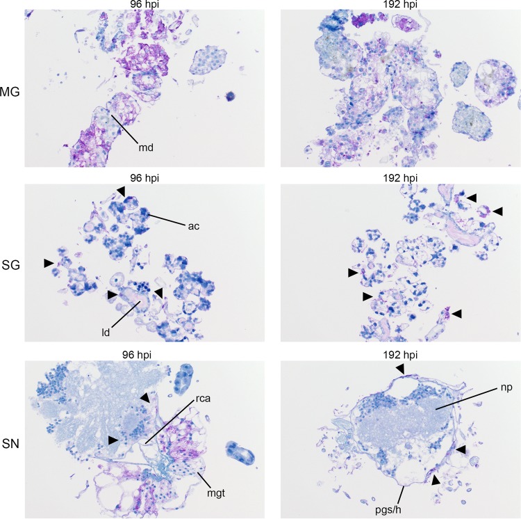
FIG 6 .
Progression of POWV protein synthesis in infected organ cultures. Magnification, ×20 (POWV-infected MG, salivary gland [SG], and synganglion [SN]). md, midgut diverticulum; ld, lobular duct; a.c., acinus; pg/h, periganglionic sinus/sheath; n.p., neuropile; rca, retrocerebral area; mgt, midgut tissue. POWV proteins within tissue are denoted by the prominent purple color. Mock-infected organs at 192 hpi were processed and stained in the same manner, using POWV antibody to identify any potential nonspecific staining background (see Fig. S3).

