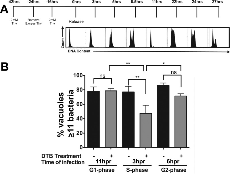
FIG 5 .
Legionella pneumophila intracellular replication is diminished in S phase HeLa cells. (A) HeLa cells were synchronized by a double-thymidine block (DTB [−42 to 0 h]), released, and challenged with L. pneumophila at 3, 6, and 11 h postrelease (Materials and Methods). Histograms represent the cell cycle profile of Hoechst-stained cells at the indicated times postrelease (hpr). (B) L. pneumophila cells show defective growth in S phase cells. Lp01 was used to challenge HeLa cells at the noted time points, and the noted cell cycle phases were determined based on flow analysis. Cells were fixed and permeabilized 14.5 h after challenge and stained with anti-Legionella, and the numbers of bacteria per vacuole were scored microscopically, displaying the number of vacuoles having more than 11 bacteria. ns, not significant; *, P ≤ 0.05, and **, P < 0.01, by one-way ANOVA with post hoc Bonferroni’s multiple-comparison test.

