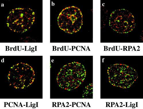
Figure 5.
Etoposide-induced RPA2 foci do not colocalize with replication factories. HeLa cells were treated for 1 h with 100 μM VP-16. Cells were stained with the polyclonal antibody to hLigI (a), with the polyclonal antibody to PCNA (b), and with the 9H8 mAb to RPA2 (c). Antibodies were revealed with a Cy5-conjugated secondary antibody (red). In the same panels sites of BrdU incorporation were revealed by the FITC-conjugated anti- BrdU mAb (green). Confocal laser images of the same cell were taken and merged. Yellow spots indicate the extent of protein-BrdU colocalization. d, cells were costained with the PC10 mAb to PCNA (green) and the polyclonal antibody to hLigI (red). e, cells were costained with the 9H8 mAb to RPA2 (green) and the polyclonal antibody to PCNA (red). f, cells were costained with the 9H8 mAb to RPA2 (green) and with the polyclonal antibody to hLigI (red). mAbs were revealed with the FITC-conjugated anti-mouse secondary antibody, and polyclonal antibodies were revealed with the Cy5-conjugated anti-rabbit secondary antibody. Confocal laser images of the same cell were taken and merged. Yellow spots indicate the extent of colocalization between the different proteins.

