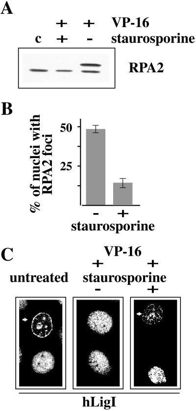
Figure 7.
Staurosporine prevents the phosphorylation of RPA2 and the redistribution of RPA2 and hLigI. (A) Western blot analysis of 0.5% Triton X-100–insoluble cell extracts prepared from HeLa cells treated for 1 h with 100 μM VP-16 either in the presence (+) or in the absence (−) of 10 μM staurosporine. Staurosporine was added to the medium 15 min before VP-16. Untreated cells (c) were also analyzed. RPA2 was revealed with 9H8 mAb. (B) The histogram indicates the percentage of cells with RPA2 foci after a treatment of 1 h with VP-16 (100 μM), either in the absence (−) or in the presence (+) of 10 μM staurosporine. Error bars indicate the average error of the mean as determined from three separate experiments. (C) Immunofluorescence analysis of the subnuclear distribution of hLigI in untreated HeLa cells or in cells grown for 2 h in 100 μM VP-16–containing medium either in the absence (−) or in the presence (+) of 10 μM staurosporine. Cells were stained with 2B1 mAb and with FITC-conjugated goat anti-mouse IgG secondary antibody. Arrows point to cells in which hLigI has a typical mid-S-phase pattern.

