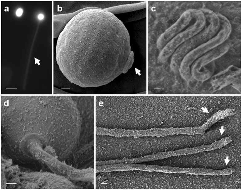
Figure 4.
Light microscope and SEM images of polar capsules and tubules. (a) Intact and activated polar capsules, pre-stained with Acridine orange. The arrow marks the florescence-stained tubule of the activated polar capsule; note that the activated capsules retain the dye. Bar = 5 μm. (b) SEM image of an unfired capsule. The operculum (arrow) is distinct. (c) Cryo-SEM image of the inverted, highly-packed tubule folded within an intact capsule. (d) Activated capsule with the everted tubule. (e) Released tubules with hooked ends (arrowed). Scale bars (b–e) 200 nm.

