Figure 1.

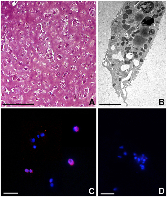
Phenotype analysis of cells recruited into the matrigel sponges by HmAIF-1 (A,B) and proliferation assay (C,D). After 1 week in vivo, MG is infiltrated by numerous cells stained by crystal violet and basic fuchsine (A). Ultrastructural analysis at TEM (B) confirm the macrophagic nature of the migrating cells in the MG sponges. Immunofluorescence experiment (C) shows BrdU+ nuclei (in red) in most of the cell migrated in the MG sponges. No signal is detected in negative control experiments (D) where the primary antibody was omitted. Nuclei are counterstained with DAPI (blue). Bar in (A) 50 µm; bar in (B) 2 µm; bars in (C,D) 10 µm.
