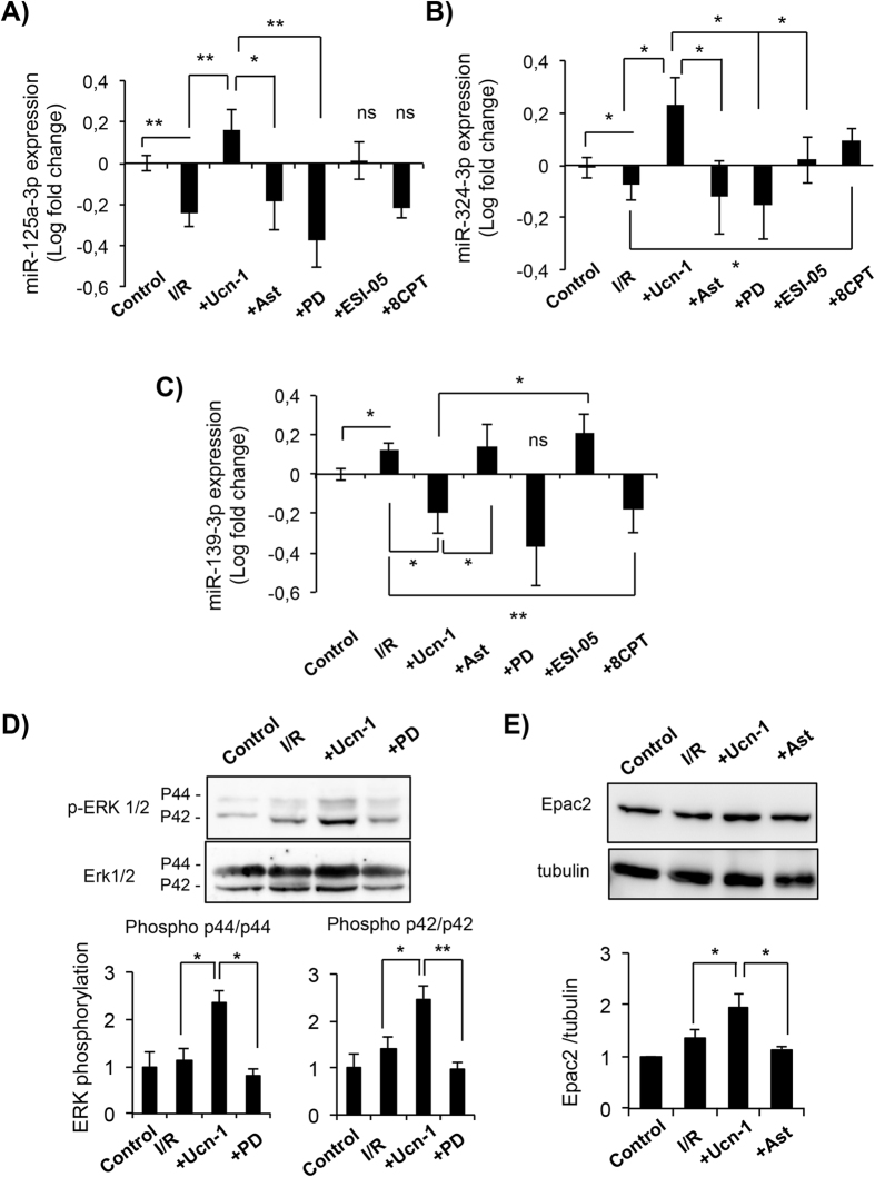
Figure 4.
Urocortin-1 regulates the expression of miRNAs in isolated cardiac myocytes through ERK1/2 and Epac2 activation. (A–C) Bar graphs show changes in the expression of miR-125a-3p (A), miR-324-3p (B) and miR-139-3p (C) in isolated cardiac myocytes treated with Ucn-1 (10 nM) in reperfusion. (D) Western blot and summary data showing ERK 1/2 phosphorylation in cells exposed to I/R (30 minutes/10 minutes). (E) Western blot and summary data showing Epac2 examined in cells exposed to I/R (30 minutes/18 hours each). “I/R” is for cells undergoing I/R protocols. “+Ast” is for cells subjected to I/R pre-treated 10 minutes with 0.5 µM astressin to inhibit CRF-R2 before the addition of Ucn-1. “+PD” is for cells pre-treated 10 minutes with 5 µM PD 098059 to inhibit ERK1/2, before the addition of Ucn-1. “+ESI-05” is for cells pretreated 10 minutes with ESI-05 (10 µM) to inhibit Epac2, before the addition of Ucn-1. “+8CPT” is for cells treated with 10 µM 8CPT at the onset of reperfusion. Values are shown in logarithmic scale and are means ± S.E.M, (n = 6–9 independent cells preparation). “*” and “**” indicate significance at p < 0.05 and p < 0.01.

