
Figure 6.
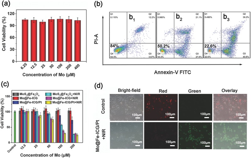
a) In vitro cytotoxicity of Mo@Fe‐ICG against L929 cells after 24 h incubation. b) Flow cytometry analysis of Hela cells under different 808 nm NIR irradiation power densities (b1: 0 W cm−2; b2: 0.5 W cm−2; b3: 1.0 W cm−2) after incubated with Mo@Fe‐ICG/Pt (100 × 10−6 m of Mo). c) In vitro cytotoxicity of MoS2@Fe3O4, MoS2@Fe3O4+NIR, Mo@Fe‐ICG, Mo@Fe‐ICG+NIR, Mo@Fe‐ICG/Pt, and Mo@Fe‐ICG/Pt+NIR against HeLa cell after 24 h incubation. The cells were either exposed to 808 nm laser (1.0 W cm−2) for 5 min or not. d) JC‐1 probes were used as the mitochondrial membrane potential indicator through comparing the fluorescence intensity ratio of red and green. The incubation concentration of Mo@Fe‐ICG/Pt is 100 × 10−6 m and the 808 nm NIR irradiation power density is 1.0 W cm−2.