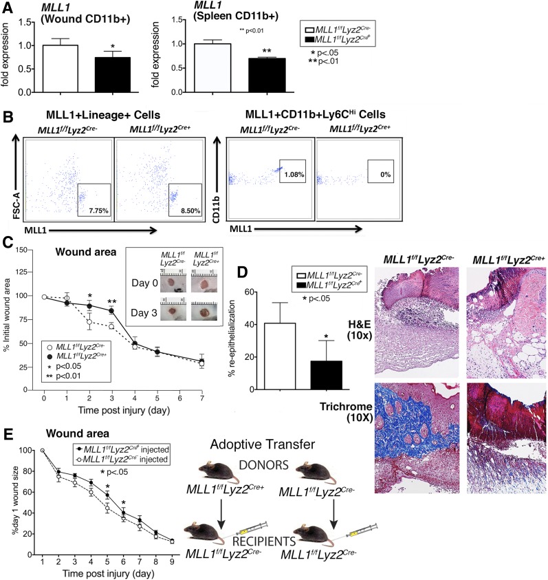
Figure 2.
Wound healing is impaired in macrophage-specific MLL1-deficient mice (Mll1f/fLyz2Cre+). We generated mice deficient in Mll1 in cells of myeloid lineage with lysosomes (monocytes, macrophages, granulocytes) by using the Cre-lox system. A: Myeloid depletion of Mll1 was examined by qPCR in MACS splenic and wound macrophages CD11b+[CD3−CD19−Ly6G−] from Mll1f/fLyz2Cre+ mice and littermate controls (Mll1f/fLyz2Cre−) (n = 10, repeated one time). B: Depletion of MLL1 was examined by flow cytometry in nonmyeloid Lin+ cells (CD3, CD19, NK1.1, Ter-119)+ and CD11b+, Ly6CHi cells from spleens of Mll1f/fLyz2Cre+ mice and littermate controls (Mll1f/fLyz2Cre−). C: Wounds were created by 4-mm punch biopsy on the backs of Mll1f/fLyz2Cre+ mice and littermate control mice. The change in wound area was recorded daily with ImageJ software until complete healing was observed. Representative photographs of the wounds of Mll1f/fLyz2Cre+ mice and littermate controls on days 0 and 3 postinjury are shown (n = 20, repeated three times). D: Wounds were created by 4-mm punch biopsy on the backs of Mll1f/fLyz2Cre+ mice and littermate control mice. Wounds were harvested on day 2, paraffin embedded, and sectioned. Sections (5 μmol/L) were stained with hematoxylin-eosin (H&E) and Masson’s trichrome. Percent reepithelialization was calculated by measuring the distance traveled by epithelial tongues on both sides of the wound divided by total distance for full reepithelialization. Representative images are shown (n = 10, repeated one time). E: CD3−CD11c−CD19−Ly6G−NK1.1−CD11b+ single-cell suspensions were isolated from Mll1f/fLyz2Cre+ and Mll1f/fLyz2Cre− spleens by MACS. Cells (1 × 10⁶) were injected intravenously in wounded (day 1) Mll1f/fLyz2Cre− mice, and wound closure was measured daily with ImageJ software (n = 15). Statistical analysis was by Student t test. Data are mean ± SEM. FSC-A, forward scatter area.

