
Fig. 7.
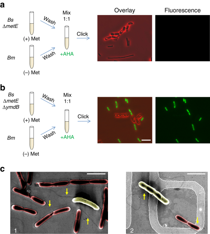
ymdB is required for Met extraction. a, b Bm (OS2) was pre grown in the absence of Met (50 μg/ml), washed in PBS×1, mixed with Bs (LS5: ΔmetE) (a) or Bs (OS21: ΔmetE, ΔymdB) (b), and resuspended in S7 supplemented with AHA (1 mM). Cells were incubated for additional 1.5 h, underwent click reaction and visualized by fluorescence microscopy. Shown are overlay images of fluorescence from AHA (green) and phase-contrast (red; left panels), and AHA fluorescence alone (right panels). Fluorescence images were normalized to the same intensity range. Scale bar represents 10 µm. c Bs (GB168: ΔymdB, Δhag, amyE::Phyperspank-ymdB) and Bm (OS2) cells were incubated at low density in LB medium for 1 h, and then fixed and visualized by HR-SEM without coating. Shown are HR-SEM images (×15,000) of (1) a single Bm cell (yellow) linked via nanotubes to several Bs cells (red), and (2) a single nanotube linking neighboring Bs and Bm. Arrows point to nanotube structures. Scale bars represent 3 µm