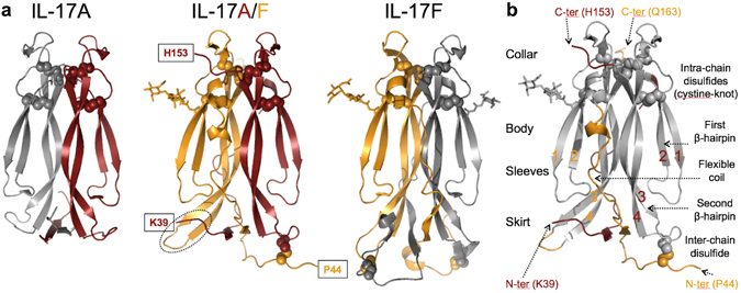
Figure 1.

Structure of human IL-17A/F. (a) Ribbon diagram of IL-17A/F (center view; A chain, carmine; F chain, orange) and comparison with IL-17A (left, 4hr9.pdb) and IL-17F (right, 1jpy.pdb). Disulfides are represented with spheres. Sugar residues are shown in stick representation. Observed N- and C-termini of IL-17A/F are boxed. The tip of the second β-hairpin of the F-chain is highlighted with a dotted ellipse. (b) Upside down orientation emphasizing the analogy between the IL-17 fold and a garment. Structurally-conserved regions in IL-17A/F with respect to the corresponding homodimers are in grey, while non-conserved structural elements are in carmine (A chain) or orange (F chain). The β-strands forming the two β-hairpins of each subunit are numbered sequentially 1 to 4.
