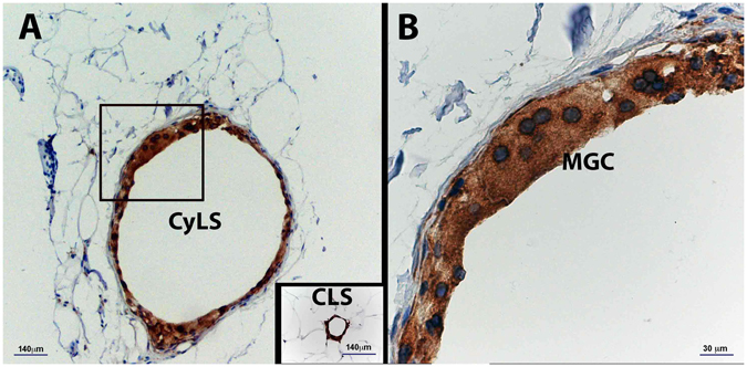
Figure 3.

Light microscopy of subcutaneous adipose tissue (SAT) of an obese patient with type 2 diabetes. Immunohistochemistry for CD68. (A) Cyst-Like-Structure (Cys-LS) formed by CD68 immunoreactive macrophages and multinucleated giant cells (MGC) surrounding fat, amidst hypertrophic adipocytes. Note the giant size of Cys-LS in comparison with hypertrophic adipocytes and the largest CLS found in the same patient. (B) Magnification of the inset in A.
