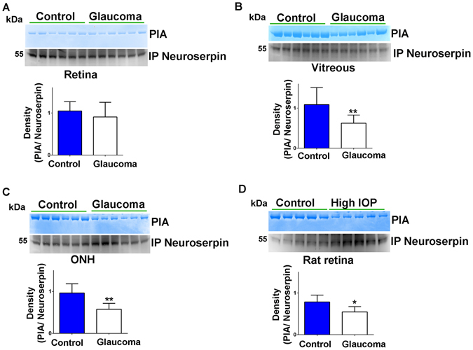
Figure 5.

Neuroserpin was immunoprecipitated from (A) human retinal (B) vitreous and (C) ONH tissue lysates using anti-neuroserpin antibody and subjected to gelatin zymography to evaluate the plasmin inhibitory activity of the neuroserpin (n = 6 each). (D) Immunoprecipitated neuroserpin from control and high IOP rat retinas were subjected to gelatin gel zymography to assess its plasmin inhibitory activity (n = 5). Immunoprecipitates were also loaded for western blotting and developed for neuroserpin immunoreactivity in each case. Blots were cropped to show the relevant band. Relative band intensities were quantified and data analysis indicated significantly decreased plasmin inhibitory activity in human vitreous and ONH glaucoma samples and high IOP induced rat retinas compared to the respective controls (*p < 0.05; **p < 0.01).
