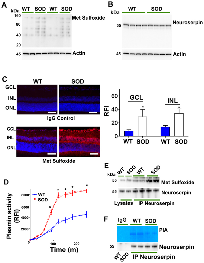
Figure 9.

Retinal lysates from WT and SOD mutant mice were immunoblotted and developed for (A) methionine sulfoxide and (B) neuroserpin reactivity (n = 4). Actin was used as internal control for normalisation in each case. (C) Increased met sulfoxide immunofluorescence (red) was observed in the retinal sections from SOD mutant mice and results compared with wild type animals and also with the sections incubated with control IgG antibodies (Dapi- blue). The relative fluorescence intensities for met sulfoxide reactivity in WT and SOD sections were quantified in the GCL and INL using ImageJ and data plotted (Scale 50 µm, n = 4, *p < 0.05). (D) Plasmin proteolytic enzymatic assay was carried out (0–300 minutes) from retinal lysates obtained from WT and SOD mutant retinas and plotted (n = 4). Dotted lines represent sigmoidal allosteric regression analysis (*p < 0.05). (E) Neuroserpin immunoprecipitates from WT and SOD mutant mice retinas along with retinal lysates were subjected to western blotting for met sulfoxide immunoreactivity. (F) Following immunoprecipitation from WT and SOD mutant mice retinal lysates with anti-neuroserpin antibody, immunoprecipitates were subjected to gelatin in-gel zymography for plasmin proteolytic inhibitory activity assay. Non-immune IgGs were used as control for immunoprecipitation.
