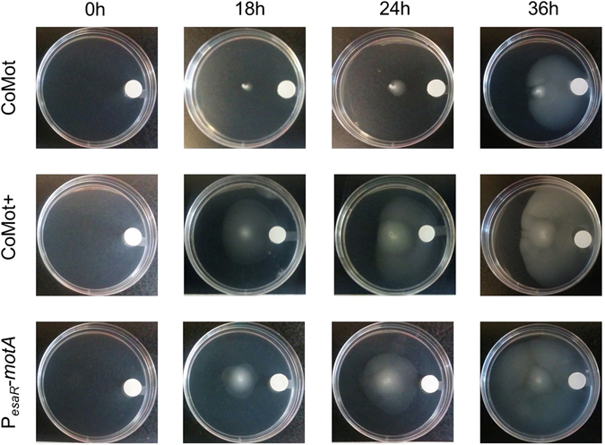
Figure 2.

CoMot and CoMot+ cells in a 3OC6HSL gradient show directional movement towards the 3OC6HSL source: A 3OC6HSL gradient was established by adding 0.02 μmoles of 3OC6HSL to a Whatmann membrane and allowing it to diffuse for 8 h. 1 μM of 3OC6HSL would be the final concentration if 0.02 μmoles of 3OC6HSL diffused uniformly through the plate. CoMot, CoMot+ or cells that constitutively express motA (∆motA transformed with a plasmid containing PesaR-motA) were then inoculated at the centre of the plate. Images were obtained following 0, 18, 24 and 36 h of incubation at 30 °C. The assay was run in triplicate for each strain and representative images are shown.
