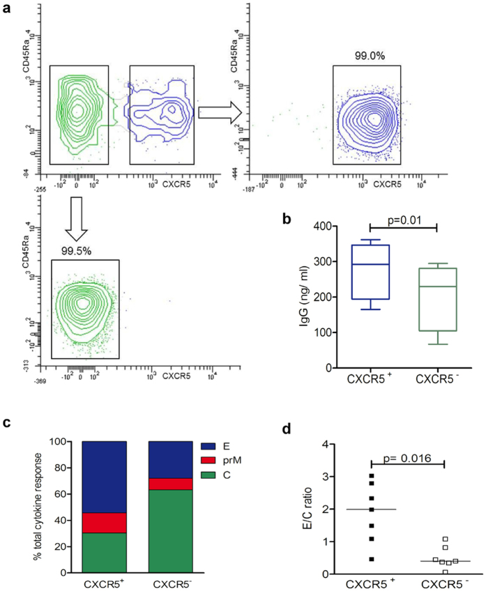
Figure 2.
CXCR5+ and CXCR5− CD4 T cell response to YF virus C, prM and E peptides. (a) CD4+ CD45Ra− memory T cells from PBMC of healthy donors were sorted into CXCR5+ (upper right) and CXCR5− cells (lower left). (b) IgG concentration revealed by ELISA in supernatants of sorted autologous B cells cultured for 10 days with either CXCR5+ or CXCR5− cells in 4 independent experiments. Statistical signifiance was determined with the two-way ANOVA. (c) Percentage of cytokine events in sorted CXCR5+ and CXCR5− subsets contributed by C, prM and E peptides as determined by intracellular cytokine staining. (d) Ratios of results with E and C peptides obtained in individual donors. Statistical signifiance was determined with the Wilcoxon matched-pairs signed rank test.

