
Figure 2.
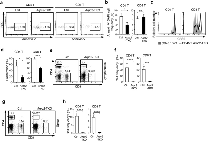
The loss of Arpc2 leads to impaired T cell homeostasis. (a) Survival was assessed in naïve T cells using flow cytometry analysis with Annexin V and DAPI staining. (b) The percentage statistics for Annexin V of naïve T cells obtained from Ctrl and Arpc2-TKO mice are illustrated. (n = 3). (c) Naïve CD4+ or CD8+ T cells isolated from CD45.2+ Arpc2-TKO mixed equally with CD45.1+ congenic Ctrl mice were labeled with carboxyfluorescein succinimidyl ester (CFSE) and co-transferred into Rag1 −/− mice. After 7 days the mice were sacrificed, and the lymphocytes were analyzed. (d) The percentage statistics for CFSE dilution proliferated CD4+ and CD8+ T cells was analyzed (n = 3). (e-h) A mixture of equal number of CD45.1+ Ctrl and CD45.2+ Arpc2-TKO bone marrow cells was transferred into Rag1 −/− mice. Flow cytometry analysis of CD4 and CD8 expression of T cells in (e) lymph nodes and (g) spleen. The percentage statistics for CD4+ and CD8+ T cells in (f) lymph nodes (n = 4) and (h) spleen (n = 6). The data are means ± S.D., for all panels: *P < 0.01; ***P < 0.001; ****P < 0.0001 by Student’s t-test, N.S.: no significance. All results are representative from at least three independent experiments.