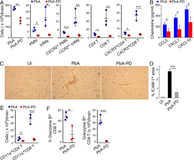
Figure 11.
MEK1/2 inhibitor treatment reduces chemotaxis-dependent brain infiltration of immune cells in malaria-infected mice. PbA-infected mice were treated i.p. with 3 mg of PD/kg body weight or vehicle daily starting at 6 h pi. A, cell numbers in the brain of PbA-infected mice at 6 days pi. The levels of PMNs and Mφs, CCR2+ Mφs, CXCR2+ PMNs, and CXCR3+ CD4+ and CD8+ T cells infiltrated to the brain are shown. B, CCL5, CXCL1, and CXCL10 chemokine levels in brain homogenates analyzed by ELISA. C, images of the sections of brains from uninfected mice (UI), PbA-infected mice (PbA), and PbA-infected and PD-treated mice (PbA-PD) at 7 days pi stained for ICAM-1. D, ICAM-1+ blood vessels in 20 microscopic fields of brain sections were counted and data plotted. E and F, brain cells were stained with antibodies against T cell markers, CD11a (LFA-1), and granzyme B. The stained cells were analyzed by flow cytometry. The gating of brain-infiltrated cells and the gating on cells expressing chemokine receptors, CD11a, and granzyme B are shown in supplemental Fig. S5. E, infiltrated CD4+ and CD8+ T cell numbers per brain. F, percentage of granzyme+ CD8+ T cells and the granzyme+ CD8+ T cell number per brain. The data are representative of three or four independent experiments; n = 3 mice/group in each experiment. Statistical analysis was performed by either one-way ANOVA with Newman–Keuls correction for multiple comparisons (D) or unpaired two-tailed t test (A, B, E, and F). Error bars, S.D. *, p < 0.05; **, p < 0.01; ***, p < 0.001.

