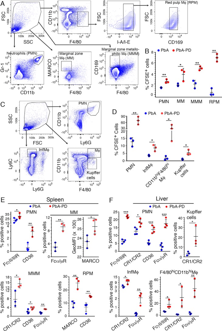
Figure 4.
Treatment with MEK1/2 inhibitor enhances phagocytic receptor expression and phagocytosis of parasites. PbA-infected mice were treated i.p. with 3 mg of PD/kg body weight or vehicle daily starting at 6 h pi. A–D, at 4 days pi, ∼2.5 × 10⁷ CFSE-labeled PbA-IRBCs were administered. After 18 h, spleen and liver cells were stained with antibodies against the indicated marker proteins and analyzed by flow cytometry. SSC, side scatter; FSC, forward scatter. A and B, spleen PMNs and Mφ subsets were gated as shown in A, and the percentage of CFSE+ cells, gated as shown in supplemental Fig. S1, was plotted (B). C and D, liver PMNs, inflammatory Mφs, and Kupffer cells were gated as shown in C, and the percentages of CFSE+ PMNs, inflammatory Mφs, CD11bhiF4/80hi Mφs, and Kupffer cells, gated as shown in supplemental Fig. S1, were plotted (D). E and F, at 5 days pi, spleen (E) and liver (F) cells were analyzed by flow cytometry. The cells were gated as shown in A and C, and the percentages of phagocytic receptor+ PMNs and the indicated Mφ subsets, analyzed as shown in supplemental Fig. S2A (spleen cells) and supplemental Fig. S2B (liver cells), were plotted. Results are representative of three (A–D) or four (E and F) independent experiments. A–F, n = 3–4 mice/group. Statistical analysis was by unpaired two-tailed t test. Error bars, S.D. *, p < 0.05; **, p < 0.01; ***, p < 0.001.

