Figure 5.
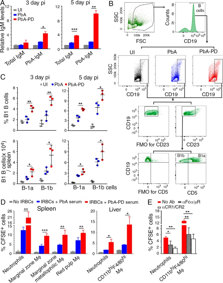
Treatment of malaria-infected mice with PD leads to increased B1 cell expansion, IgM production, and phagocytosis of parasites. A–D, PbA-infected mice were treated i.p. with 3 mg of PD/kg body weight or vehicle daily starting at 6 h pi. A, serum IgM levels (n = 4 mice/group) at 3 days and 5 days pi assessed by ELISA. B and C, flow cytometry analysis of splenic B1a and B1b B cells at 3 days and 5 days pi (n = 3 mice/group). Shown are the gating strategy and FMO control for gating CD23− cells (B) and B1a and B1b cell frequency (top) and cell numbers per spleen (bottom) in control and PD-treated mice relative to uninfected mice (C). D, the phagocytosis of IRBCs pretreated with 1:10-diluted sera from control or PD-treated mice at 5 days pi by spleen and liver PMNs and MM, MMM, and RPM from PD-treated mice at 5 days pi. Experiments were performed in triplicate. Data are a representative of four independent experiments; n = 3 mice/group in each experiment. A, C, and D, statistical analysis was by one-way ANOVA with Newman–Keuls correction for multiple comparisons. Error bars, S.D. E, the phagocytosis of CFSE-labeled IRBCs pretreated with 1:10-diluted sera from PD-treated mice at 5 days pi by liver PMNs and CD11bhiF4/80hi Mφs from PD-treated mice at 5 days pi treated with either anti-Fcα/μR antibody or anti-CR1/CR2 antibody. PMNs and CD11bhiF4/80hi Mφs from PD-treated mice not treated with antibodies were analyzed as controls. Experiments were performed in triplicate. Data represent the mean of two independent experiments, and statistical analysis was by two-way ANOVA. Error bars, S.E. Blocking of Fcα/μR and CR1/CR2 with antibodies significantly inhibited phagocytic uptake of IRBCs. *, p < 0.05; **, p < 0.01; ***, p < 0.001.

