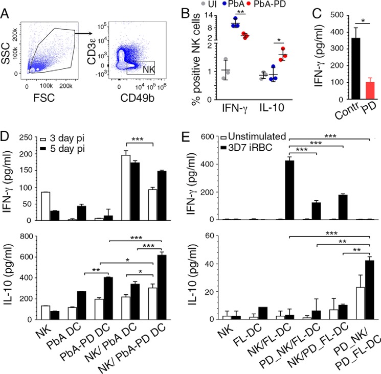
Figure 8.
MEK1/2 inhibitor treatment differentially regulates malaria-induced pro- and anti-inflammatory responses by NK cells. A and B, PbA-infected mice were treated i.p. with 3 mg of PD/kg body weight or vehicle daily starting at 6 h pi. Spleen NK cells from the infected mice at 4 days pi were analyzed by flow cytometry for cytokine production. Shown are the gating of CD49b+CD3ϵ− NK cells (A) and quantification of IFN-γ+ and IL-10+ cells (B). The results are representative of four independent experiments; n = 3 mice/group in each experiment. C, PD-treated and -untreated liver NK cells from uninfected mice were stimulated with IL-12 (1 ng/ml) and IL-18 (50 ng/ml), and IFN-γ production was measured by ELISA. D, cocultures of FACS-sorted spleen DCs from PD-treated and untreated infected mice at 3 days and 5 days pi and liver NK cells from uninfected mice stimulated with 2 μg/ml CpG ODN. Cytokines in the culture supernatants were analyzed ELISA. E, cocultures of FL-DCs and liver NK cells stimulated with P. falciparum IRBCs. Prior to coculturing, DCs alone, NK cells alone, or both DCs and NK cells were pretreated with PD for 1 h; cells not treated with PD were used as controls. Cytokines in culture supernatants were analyzed by ELISA. D and E, the results are representative of four independent experiments. Statistical analysis was by either one-way ANOVA with Newman–Keuls correction for multiple comparisons (B, D, and F) or unpaired two-tailed t test (C). Error bars, S.D. *, p < 0.05; **, p < 0.01; ***, p < 0.001.

