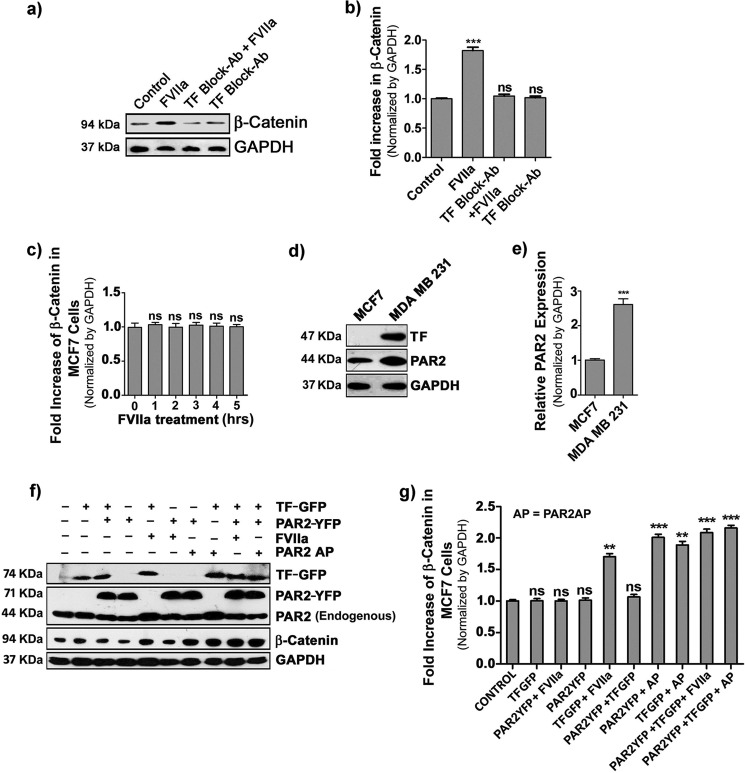
Figure 3.
Involvement of tissue factor in FVIIa-induced accumulation of β-catenin. a, MDA-MB-231 cells were treated with TF-blocking antibody (Ab) for 2 h followed by challenging with FVIIa, and β-catenin accumulation was analyzed after 4 h by Western blot analysis. b, quantification of β-catenin accumulation was performed using ImageJ and GraphPad Prism 5. c, quantitative estimation of β-catenin accumulation by Western blotting was checked in MCF-7 cells upon treatment with FVIIa at various time intervals (0–5 h). d, relative levels of TF and PAR2 were measured in MCF-7 and MDA-MB-231 cells by Western blot. e, relative PAR2 band intensity was quantified over GAPDH in MCF-7 and MDA-MB-231 cells using ImageJ and GraphPad Prism 5. f, TF, PAR2, and β-catenin levels in control, TF-GFP-, and PAR2-YFP-overexpressing MCF-7 cells were analyzed by Western blotting after FVIIa or PAR2AP treatment. g, band intensity of β-catenin over GAPDH in control, TF-GFP-, and PAR2-YFP-overexpressing MCF-7 cells was quantified using ImageJ and GraphPad Prism 5. Error bars represent ±S.E. of the mean. **, p < 0.05; ***, p < 0.001; ns, non-significant using Student's t test; n ≥ 3. AP, agonist peptide.

