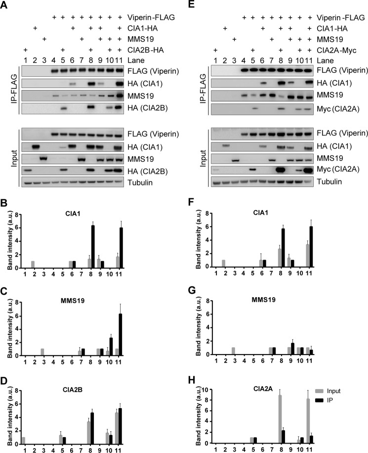
Figure 3.
CIA1 and CIA2B mediate the indirect interaction of MMS19 with viperin. A and E, HEK293T cells were transiently transfected with plasmids encoding FLAG-tagged viperin and tagged (HA or Myc) or non-tagged CIA-targeting factors as indicated (+). Anti-FLAG–viperin immunoprecipitation and sample analysis by immunoblotting were performed similar to Fig. 2. Tubulin staining served as reference. The chemiluminescence associated with CIA1-HA (B and F), MMS19 (C and G), CIA2B-HA (D), or CIA2A-Myc (H) was quantified. The signal intensity of input samples was normalized to tubulin (gray bars), and the signals associated with the immunoprecipitated samples were normalized to immunoprecipitated viperin, which was normalized to both the amount of tubulin and viperin in the total cell lysates (black bars, mean values ± S.D.; n = 3).

