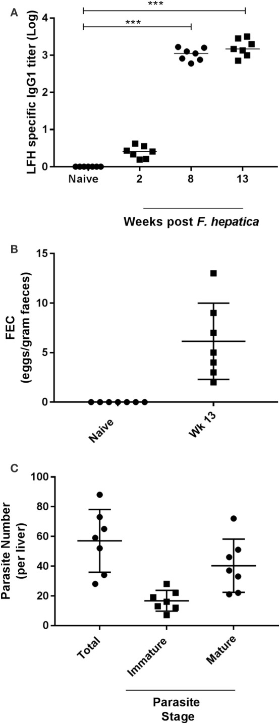
Figure 1.

Parasitological parameters and seroconversion of infected animals. (A) Animals were examined for pre- and postinfection parasite-specific IgG1 antibody titers. Using a capture ELISA endpoint, log antibody titers were determined for each animal at the indicated time points. (B) Fecal egg counts (FEC) were performed using 1 g of feces from each animal before (naïve) and after infection at week 13. Data are presented as eggs per gram of feces. (C) At postmortem, livers were removed and examined from each animal. Total parasite numbers were determined for each liver. Additionally, parasites were classified as being immature or mature for each animal also. Each data point presented represents and individual animal with medias for each time point shown on the graph; data in (A) were examined for significance using a Kruskal–Wallis with Dunn’s multiple comparisons where ***P < 0.001.
