Figure 3.
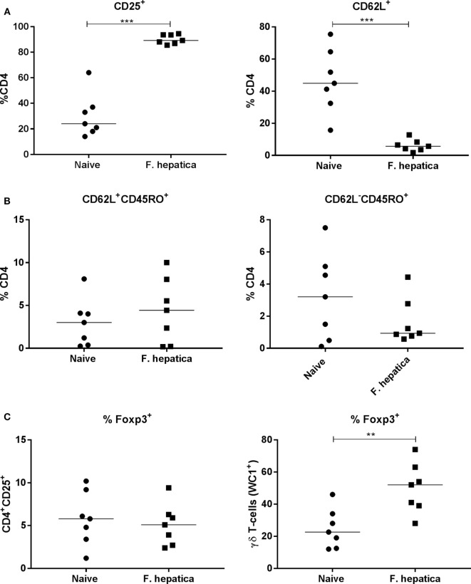
Activation, memory, and regulatory characteristics of the CD4 T-cell pool from infected lymph nodes. Single cell suspensions from the hepatic lymph node of week 13 Fasciola hepatica-infected animals (squares) or naïve controls (circles) were stained for CD4, CD25, CD62L, WC1, CD45RO, or intracellularly for Foxp3. (A) Shows an increase in the percentage of CD4+CD25+ cells, left, and a concomitant decrease in CD4+CD62L+ double-positive cells, right, indicating the presence of an activated CD4 population. (B) Staining for central memory T-cells (left) and effector memory T-cells (right) revealed that there was no significant change in the proportion of either of these populations although a noted wide variation was present in both naïve and infected animals. (C) Expression of the regulatory marker, Foxp3, was not changed in the CD4+CD25+ population when compared in naive and infected animals (left graph), but was found to be significantly upregulated in the γδ (WC1+) population between the two groups (right graph). Each point represents an individual animal. Significant differences between populations are indicated on the graphs determined using a Mann–Whitney U-test; **P < 0.01, ***P < 0.001.

