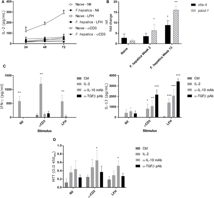
Figure 4.
Failure to produce IL-2 and markers of anergy are coincident with non-responsive CD4 T-cells. (A) CD4 T-cells were isolated from the hepatic lymph node (HLN) of naïve or infected animals and cultured with α-CD3, LFH, or PBS (nil) for 72 h. Supernatants were recovered at every 24 h and tested for IL-2. There is a clear failure of CD4 T-cells from the HLNs of infected animals to secrete IL-2. However, naïve CD4 T-cells readily secrete IL-2 over time with a near linear accumulation of cytokine. (B) mRNA from CD4 T-cells was isolated, cDNA prepared, and qPCR carried out. There is a clear increase in pdcd1 expression from week 2 onward when compared to naïve animals. Ctla4 expression is significantly upregulated at week 13 but not week 2. (C) The effect of exogenous IL-2, or neutralizing IL-10 or TGF-β antibody on the expression of IFN-γ (left) and IL-13 (right) in infected CD4 T-cells from HLN was examined. There was a clear impact of IL-2 on IFN-γ, and IL-13 production was restored in the presence of all three treatments but to varying degrees. Ctrl represents cells treated with isotype Ab only at equal concentrations to neutralizing Ab. (D) CD4 T-cell proliferative responses in the HLN were assessed using the MTT dye assay after stimulation in the presence of treatments as in (C). LFH and α-CD3 responsiveness was restored in the presence of α-IL-10 when compared within stimulation to non-treated controls. Bars represent the mean of seven animals ±SEM. Significant differences were determined using Friedman test with a post hoc comparison using a Wilcoxon ranked-sign test (A,C,D) and Kruskal–Wallis with Dunn’s multiple comparisons (B) where *P < 0.05, **P < 0.01, ***P < 0.0001.

