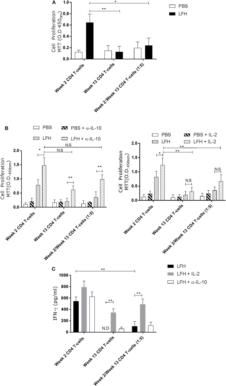
Figure 5.
Anergic CD4 T-cells are suppressive toward autologous responsive counterparts. CD4 T-cells taken from animals’ 2 weeks of postinfection were cocultured with autologous CD4 T-cells isolated from the hepatic lymph node at postmortem. (A) Proliferation in response to LFH is still detectable in week 2 CD4 T-cells, as per previous findings; however, when anergic week 13 CD4 cells are added at a ratio of five cells per 1 week 2 responder cells, we see a strong suppression of proliferation. (B) A similar coculture was established and the effects of α-IL-10 (left graph) and exogenous IL-2 (right graph) was determined. In the presence of α-IL-10, there was significant reversal of the negative effects of anergic CD4 cells on their responsive counterparts; however, there was no positive effect of exogenous IL-2. (C) IFN-γ levels in LFH-stimulated cells untreated (isotype control) or treated with IL-2 and α-IL-10 were determined in the coculture system. In the presence of week 13 anergic CD4 cells, the IFN-γ response, from responsive week 2 cells, was suppressed while IL-2, but not α-IL-10 treatment, could abrogate the effect of week 13 CD4 T-cells on the responsive population. Bars represent the mean of seven animals’ ±SEM. Data in (A) and (B,C) were tested by Kruskal–Wallis with Dunn’s multiple comparisons and Friedman test with a post hoc comparison using a Wilcoxon ranked-sign test, respectively. ND, not detected; NS, not significant, *P < 0.05 and **P < 0.01.

