Figure 8.
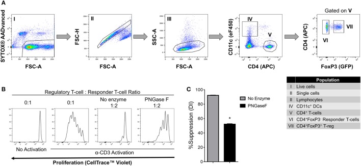
Suppression of CD4+ responder T-cell proliferation by PNGase F-treated Treg in the presence of dendritic cells (DC). (A) Gating strategy for purification of CD11c+ DCs, CD4+ Tconv, and Treg from C57BL/6 FoxP3.EGFP mouse spleen and lymph nodes. Individual sorted populations are identified by Roman numerals and described in the text box. (B,C) Fluorescence-activated cell sorting (FACS)-purified CD4+ Tconv were cocultured in the presence of FACS-purified CD11c+ DCs without Treg (0:1) and with no enzyme or PNGase F-treated Treg at Treg: responder T-cell ratio 1:2 for 4 days with anti-CD3 stimulation. (B) Proliferation of CellTrace™ Violet-labeled CD4+ Tconv analyzed by flow cytometry at the end of the culture period. (C) Graph of the suppressive potency of no enzyme and PNGase F-treated Treg on CD4+ Tconv. Suppressive function was quantified based on Tconv division index (DI) and presented as the calculated percent suppression [%Suppression (DI)]. Data represent mean ± SD (n = 3 technical replicates). The experiments shown in panels (B,C) are representative of three independent experiments with consistent results. Statistical analysis was performed by permutation test with an unpaired design (*p value ≤ 0.1).

