
Fig. 1.
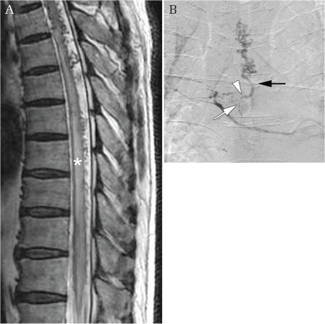
Dural arteriovenous fistula (AVF). (A) Sagittal thoracic image using T2-weighted MR imaging showing congestion of the spinal cord (asterisk). (B) An antero-posterior angiogram of the thoracic intercostal artery showing feeding meningeal arteries (white arrow), the fistula (white arrowhead), and draining vein (black arrow).