
Fig. 3.
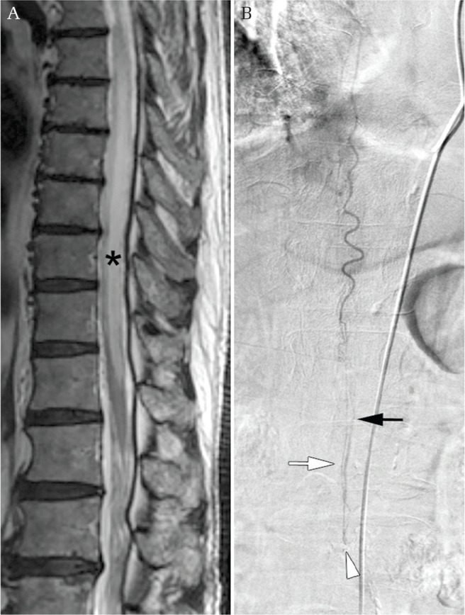
Intradural perimedullary AVF subtype I (a single feeder, a single small AVF). (A) A sagittal thoracic image using T2-weighted MR imaging showing congestion of the spinal cord (asterisk). (B) An angiogram of the thoracic intercostal artery showing intradural perimedullary AVF subtype I (arrowhead) fed by the filum artery (white arrow), and draining vein (black arrow).