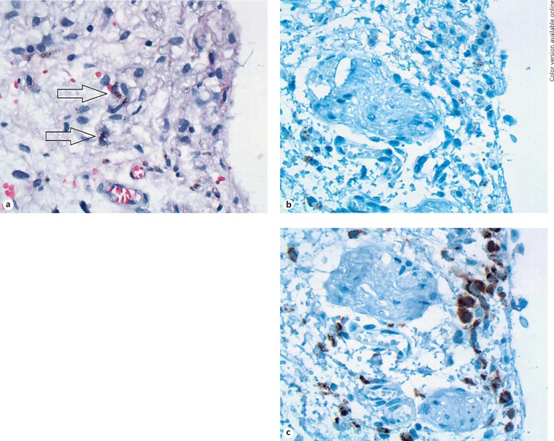
Fig. 4.
Histopathology. a Histology of the pigmented lesion showing conjunctival stroma with scattered cells with brown cytoplasmic pigment and small nuclei (arrows). HE stain. Original magnification, ×40. b The cells are not reactive for Melan-A. In some cells, brown pigmentation persisted despite melanin depigmentation, but it is very distinct from the immunohistochemical reaction product. Melan-A stain after melanin depigmentation. Original magnification, ×40. c The cells show positive staining for macrophage-specific CD68. CD68 staining after melanin depigmentation. Original magnification, ×40.

