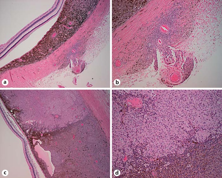
Fig. 2.
Histopathology. a Low magnification of choroidal tumor (H&E, ×4). b Extrascleral extension along ciliary artery (H&E, ×20). c Tumor heterogeneity (H&E, ×20) with eosinophilic appearance of the epithelioid portion of the tumor in comparison to the spindle B cell portion of the tumor. d High-magnification view of tumor heterogeneity (H&E, ×40).

