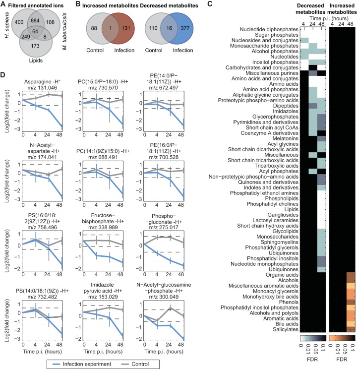
FIG 2 .
Analysis of changes in the host metabolome during M. tuberculosis infection. (A) Annotation statistics of all detected ions after the filtering procedure. (B) Comparison of the number of changing metabolites in control and infection samples at 48 h p.i. defined using a fold change (FC) cutoff of |log2 FC| > log2 1.5 and FDR < 0.05. (C) Chemical class enrichment analysis of changing metabolites at each time point. Metabolites were displayed only if the enrichment false-discovery rate (FDR) was <0.1. (D) Temporal profiles of metabolites representative of certain metabolite classes. Mean fold change values of at least three independent experiments are shown for infection and control experiments with 95% confidence intervals (t test). The dashed lines correspond to |log2 FC| = log2 1.5) (data from three biological replicates and two technical replicates). PC, phosphatidylcholine; PE, phosphatidylethanolamine; PS, phosphatidylserine.

