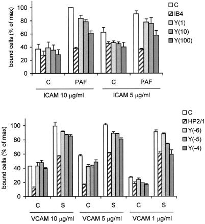
Figure 6.
Adhesion of eosinophils to ICAM-1 or VCAM. Eosinophils were calcein-loaded, incubated for 30 min with Y27632 at the indicated concentrations (between brackets, in μM), or with neutralizing antibodies against α4- (HP2/1) or β2- (IB4) integrins. Then the cells were placed in 96-well plates coated with ICAM-1 (5–10 μg/ml) or VCAM (0.1–5 μg/ml) containing control buffer or serum-containing buffer and incubated for 5 min at 37°C. The cells were washed and lysed and fluorescence intensity was measured. The results are expressed as percentage of maximal binding (mean ± SEM of 2–5 experiments performed in duplicate).

