
Figure 6.
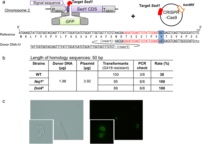
Direct fusion of GFP-coding sequence into a chromosomal locus. (a) Schematic showing integration of the GFP-coding fragment into the Sed1 CDS. The CRISPR-Cas9 plasmid targeted (indicated by lightning bolts) the end of the signal peptide-coding region of Sed1. Dashed boxes indicate homologous regions. Left arm is homologous to the end of to the signal peptide-coding sequence (bp 8 to 57 with respect to the start of the Sed1 coding sequence). Right arm is homologous to Sed1 sequence (bp 58 to 107 with respect to the start of the Sed1 coding sequence) with substitution at bp 84 (G to A; indicated by lower-case letter in red font) to eliminate a PAM. (b) The successful integration rate was calculated as PCR check * 100. (c) Representative microscopic image of positive transformants (Nej1°/GFP_Sed1) under bright-field (left) and fluorescent (right) illumination. Insets show 3-fold magnified images.