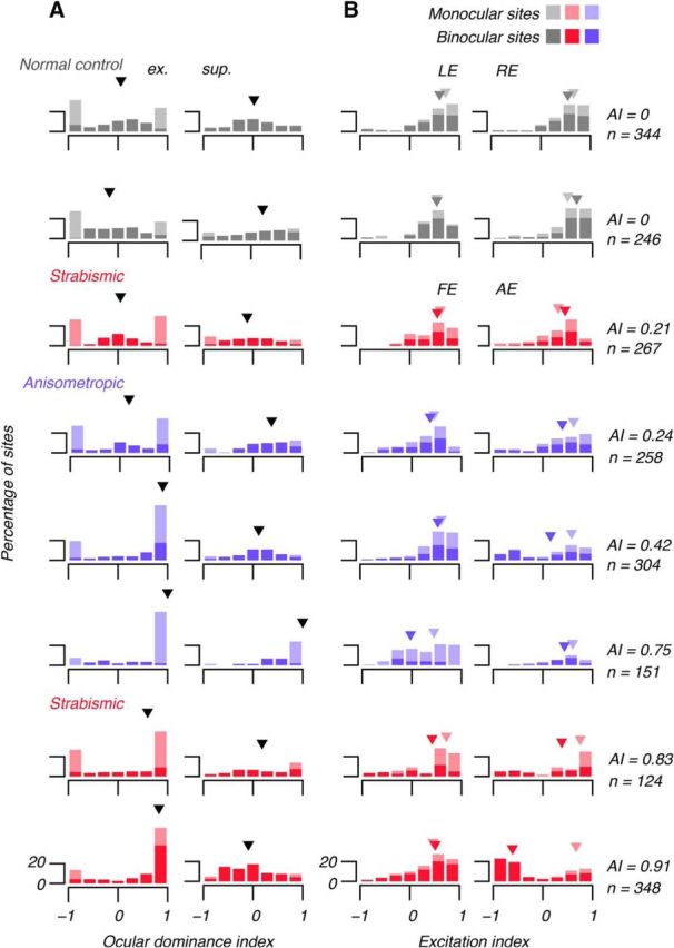
Figure 7.

Histograms showing all subjects' ODIs and EIs. Left column of A shows ODIe computed using excitatory (ex.) components of RFs (RFs) (e.g., Fig. 6). Right column of A shows ODIs computed using RF suppression (sup.). In severe amblyopia, ODIe tended to 1, indicating FE domination. There was no systematic effect on ODIs (arrowheads show medians). Left column of B shows EI in left (normal eyes) and FE (AE). Right column of B shows EI in REs and AEs. Dark arrowheads show medians of binocular sites and light arrowheads show medians of monocular sites. There was little systematic effect of amblyopia on EI in FEs; there excitation tended to outweigh suppression (EI > 0), but amblyopia altered RF composition in the AE: at binocular sites in the AE, RF suppression outweighed excitation (EI tended to −1). Stimulation of the AE suppressed binocular cortex.
